
Musculoskeletal Ultrasound for Sports Injuries in Ajax
Learn how musculoskeletal ultrasound in Ajax looks at tendons, muscles and ligaments after a sports injury, how it differs from X-ray, and how to prepare.
· 9 min read
A weekend soccer game, a slippery step off a curb, a pickup basketball landing that went slightly wrong: sports injuries rarely announce themselves politely. Often there is a sharp moment, then a dull ache that lingers longer than expected. Maybe the shoulder that felt stiff after a summer of tennis still catches when you reach overhead, or the back of your ankle has been sore since you added hills to your running route. When a health-care provider wants a closer look at what is happening beneath the skin, one of the tools they may reach for is musculoskeletal ultrasound.
This guide explains what musculoskeletal ultrasound is, what it can and cannot show, how it fits alongside X-ray, and what to expect if your provider sends you for a scan at Rossland Diagnostic Clinic in Ajax. It is written for active people of all ages, from teenage athletes and weekend league players to retirees who walk, cycle or golf. It is general information only; your own health-care provider is the right person to decide which test, if any, suits your situation.
What musculoskeletal ultrasound looks at
“Musculoskeletal” simply means the muscles and the skeleton, along with everything that connects and cushions them. Ultrasound uses high-frequency sound waves to build a live picture of the soft tissues, and it is especially good at showing structures that sit relatively close to the surface of the body.
Areas that are commonly examined include:
- Tendons, the cords that attach muscle to bone, such as the Achilles tendon at the back of the ankle or the rotator cuff tendons in the shoulder
- Ligaments, the tough bands that connect bone to bone and help stabilize joints like the ankle, knee and elbow
- Muscles, where a provider may want to look for a strain or a tear
- Bursae, the small fluid-filled sacs that reduce friction around joints
- Fluid collections around a joint or within soft tissue
- Lumps and swellings that a provider wants to characterize
Because it shows soft tissue rather than bone in detail, musculoskeletal ultrasound answers a different set of questions from an X-ray. Understanding that difference can help the process make more sense when you are referred for one test rather than the other.
Ultrasound and X-ray: different tools for different questions
A lot of people assume that a sports injury automatically means an X-ray. Sometimes it does. X-rays are very good at showing bone, so they are often a first step when a provider is concerned about a fracture, a dislocation or a change in the joint’s bony structure. At our clinic, general digital X-ray is offered as a walk-in service during opening hours, though the department may close once daily capacity is reached, so calling ahead for live availability is wise.
Tendons, ligaments and muscles, however, do not show up well on a standard X-ray. That is where ultrasound comes in. The two tests are not competitors so much as teammates, and it is not unusual for a provider to order both: an X-ray to look at the bones and an ultrasound to look at the soft tissues around them.
A few practical differences are worth knowing:
- Radiation. Ultrasound uses sound waves, not ionizing radiation. X-ray uses a low dose of radiation.
- Motion. Ultrasound produces moving, real-time images. An X-ray captures still pictures.
- Depth. Ultrasound is best for structures relatively near the surface. Deep structures, or areas hidden behind bone, are harder for sound waves to reach.
- Comparison. With ultrasound, the sonographer can easily scan the same area on your uninjured side for comparison if that is useful.
Which test is appropriate depends on your symptoms, the provider’s examination and what they need to rule in or out. If you are unsure why one test was chosen over another, it is a fair question to ask the person who ordered it.
Why a provider might order a scan after a sports injury
Every injury is individual, and only your provider can decide whether imaging is needed. That said, musculoskeletal ultrasound is commonly requested to help assess situations such as:
- Ongoing pain around a tendon, for example at the heel, the outside of the elbow or the front of the knee
- Shoulder pain or weakness when lifting the arm, where the rotator cuff is being considered
- A suspected muscle strain or tear, such as in the calf or hamstring, that has not settled as expected
- Swelling around a joint, where the provider wants to know whether fluid is present
- An ankle or wrist sprain that remains unstable or painful longer than anticipated
- A new lump that appeared after an injury or developed over time
The scan provides information. It does not, on its own, make a diagnosis or decide a treatment plan. Your provider combines the imaging report with your history, a physical examination and sometimes other tests before discussing next steps with you.
What happens during the scan
A musculoskeletal ultrasound at our Ajax clinic is a straightforward, hands-on exam. Here is a general outline of what to expect.
Getting positioned
You will be asked to expose the area being examined. For a shoulder, that may mean changing into a gown or wearing a loose tank top. For a knee or ankle, shorts are often easiest. You will sit or lie on a padded examination table in whatever position gives the sonographer the best view.
The gel and the probe
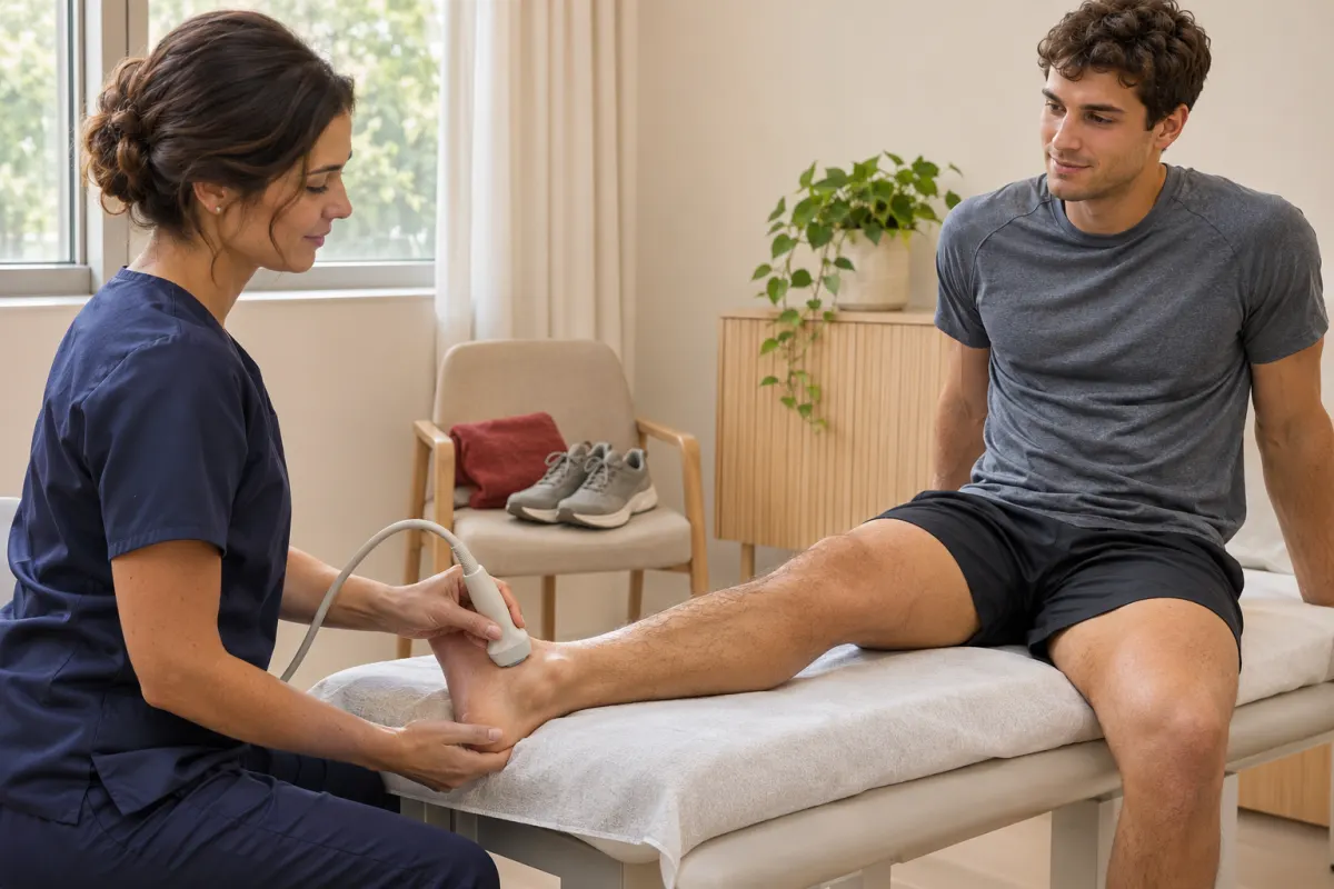
The sonographer applies a water-based gel to the skin. Gel helps the sound waves travel between the probe, called a transducer, and your body. The sonographer then moves the probe over the area, adjusting angle and pressure to bring different structures into view. Most people find this painless, though pressing over an area that is already tender can be uncomfortable. If something hurts, say so; the sonographer can adjust.
Moving during the exam
One of the useful features of ultrasound is that it can show tissues while they move. The sonographer may ask you to rotate your arm, flex your foot, bend your knee or tighten a muscle while they watch. This dynamic view can reveal how a tendon glides or how a joint behaves under gentle motion, which a still image cannot always capture.
Comparing both sides
Depending on what your provider has asked for, the sonographer may also scan the matching area on your other side. Seeing the uninjured shoulder or ankle gives a helpful point of reference for how your tissues normally look.
When the scan is finished, the gel wipes off easily and you can get dressed and go about your day. There is no recovery time from the ultrasound itself.
Preparing for your appointment
Preparation for musculoskeletal ultrasound is usually simple, but a few small steps help the visit go smoothly.
- Bring your health card and requisition. Your provider’s requisition tells the clinic which area to scan and why.
- Wear practical clothing. Choose something that lets you uncover the area easily: shorts for a leg exam, a loose top for a shoulder or elbow.
- Leave jewellery at home if it sits near the area being examined, such as rings for a hand scan or anklets for an ankle scan.
- Note your symptoms. A short list of when the pain started, what activity triggered it and what movements make it worse can help you answer questions clearly.
- Ask about specific instructions. Some ultrasound exams elsewhere in the body need fasting or a full bladder. Musculoskeletal scans generally do not, but you will receive instructions when you book, and it is always fine to call to confirm.
Our patient preparation page has exam-by-exam guidance if you have more than one appointment coming up.
What ultrasound cannot do
It is helpful to have realistic expectations. Musculoskeletal ultrasound is a valuable tool, but it has limits.
- It does not show the inside of bone. Sound waves reflect off bone surfaces, so fractures and bony changes are generally better assessed with X-ray or other imaging.
- Deep structures can be hard to see. Tissues deep within a large joint, or hidden behind bone, may not be visible.
- It is not a treatment. The scan helps your provider understand what is going on. Rest, rehabilitation, referral to a specialist or any other next step is a decision you make with your provider.
- It may not be the final test. Sometimes ultrasound answers the question completely. Other times it narrows things down and your provider decides further imaging or assessment is worthwhile.
None of this is a reason to worry. It simply means that imaging is one piece of a larger picture, and the picture is put together by the person caring for you.
Where imaging fits in getting back to activity
Most active people want one thing after an injury: to get back to what they enjoy. Imaging can support that goal by giving your provider clearer information, but it is only one part of the process.
A typical sequence might look like this:
- You notice an injury and see your family doctor, nurse practitioner, sports medicine physician or another provider.
- The provider examines you and decides whether imaging would help. If so, they complete a requisition.
- You book the scan, attend the appointment and go home the same day.
- The completed report is sent to the provider who ordered the test.
- You and your provider discuss the findings and decide together on a plan, which may include rest, physiotherapy, activity changes or further assessment.
Resist the temptation to judge your injury by how the scan felt or by anything you glimpse on the monitor. Sonographers capture the images, while interpretation and the conversation about what the results mean belong with the physicians and your own provider. Your provider is also the right person to tell you when it is reasonable to return to sport.
For families in Ajax, Pickering and Whitby juggling school sports, work and recreational leagues, having a nearby ultrasound service with Saturday hours can make that sequence a little easier to fit into a busy week. Our clinic is on Rossland Road East in Ajax, with free parking and ground-floor access, which is helpful if you are arriving on crutches or with a sore ankle.
Ready to book
If your health-care provider has given you a requisition for a musculoskeletal ultrasound, our team can help you find a time. Call us at 905-426-3111, or request an appointment online and we will contact you to confirm availability. We are open Monday to Friday from 9:00am to 5:00pm and Saturday from 8:00am to 3:30pm.