
Lumps and Bumps in Ajax: Soft Tissue Ultrasound Explained
Found a lump under the skin? Learn how soft tissue ultrasound in Ajax helps describe it, what details to note beforehand, and what the report can and can't say.
· 8 min read
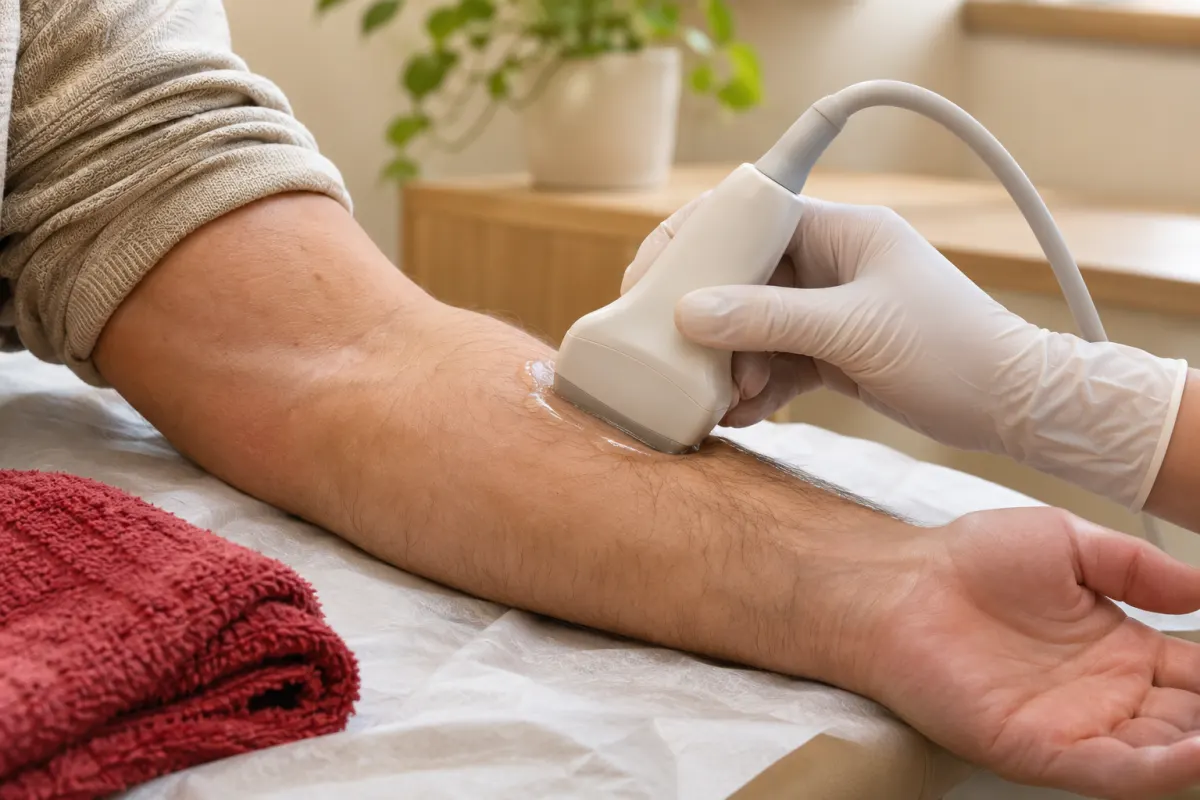
Before reading any further, take a moment with this short checklist. If you have a lump you are wondering about, these are the details your health-care provider, and later the sonographer, will find most useful:
- Where exactly is it? Note the body part and, if you can, a landmark, such as “two finger-widths above the left elbow crease.”
- When did you first notice it? An approximate date is fine.
- Has it changed? Growing, shrinking, or staying the same size.
- How does it feel? Soft or firm, movable or fixed in place.
- Is it tender, red or warm?
- Has anything happened to the area? An injury, an insect bite, a recent infection or surgery.
Writing these answers down takes a few minutes and can make both your appointment with your provider and your ultrasound more productive. The rest of this article explains why soft tissue ultrasound is such a common test for lumps, what it can tell your provider, and what to expect on the day. It is written with patients from Ajax, Pickering and Whitby in mind.
Why lumps are so often scanned with ultrasound
Most lumps and bumps under the skin are not dangerous. Many are fatty deposits, small cysts or swollen lymph nodes reacting to a minor infection. But a lump cannot be fully understood by touch alone, and that is where ultrasound helps.
A soft tissue ultrasound uses high-frequency sound waves to create pictures of the layers just under the skin: the fat, the muscle, the tendons and the small structures in between. Because these layers are close to the surface, ultrasound can show them in fine detail. It does not use radiation, it is painless for most people, and the sonographer can scan directly over the lump while you point to it.
Our article on musculoskeletal ultrasound for sports injuries covers how ultrasound looks at tendons, ligaments and muscles after an injury. This article focuses on a different question: not “what did I damage?” but “what is this thing I can feel?”
What the scan can describe
A radiologist reviewing a soft tissue ultrasound will typically comment on several features. Knowing what they are can make your report easier to follow.
Solid or fluid-filled
This is often the first and most useful distinction. Fluid-filled lumps, like many cysts, look quite different on ultrasound from solid ones.
Size and depth
The lump is measured in more than one direction. Its depth matters too: is it in the skin itself, in the fat just beneath, or deeper, within or below the muscle?
Shape and edges
Smooth, well-defined edges are described differently from irregular ones. The report may also mention whether the lump has a capsule or blends into the tissue around it.
Internal pattern
The texture inside the lump, how bright or dark it appears and whether it looks uniform, gives the radiologist further clues.
Blood flow
Using a Doppler setting, the sonographer can see whether there is blood flowing within or around the lump. Some lumps have none; others have a noticeable pattern.
Relationship to nearby structures
Is the lump attached to a tendon, a joint or a blood vessel? Is it separate from them? This can be important for understanding what it is and for planning any next steps.
Lumps that ultrasound commonly helps to characterize
Ultrasound findings are interpreted by a radiologist and discussed with you by your provider, so the list below is not a way to identify your own lump. It simply shows the range of everyday things a soft tissue scan is often used to look at.
- Lipomas. Soft, slow-growing collections of fat cells, often found on the back, shoulders, arms or thighs. They are very common.
- Skin cysts. Small lumps in or just under the skin, sometimes with a visible central point.
- Ganglion cysts. Fluid-filled lumps that often appear near the wrist or the top of the foot, sometimes changing in size.
- Lymph nodes. Small bean-shaped glands in the neck, armpit and groin that can swell when the body is fighting an infection. The scan can describe their size and shape.
- Fluid collections. A pocket of fluid after an injury or a procedure, or a swollen bursa near a joint, such as at the elbow.
- A cyst behind the knee. A fluid-filled swelling in the back of the knee, often linked to something happening inside the joint.
- Hernias. A bulge in the groin or near the belly button may be a hernia rather than a lump in the tissue; our guide to hernia ultrasound covers that scan in detail.
Lumps in the neck may be investigated with a thyroid ultrasound if they seem to involve the thyroid gland, and breast lumps follow their own pathway, so your provider will choose the right exam for the location.
When to see your provider sooner rather than later
This article is about lumps that a provider has already decided to investigate with ultrasound. If you have not yet had yours looked at, it is worth booking an appointment with your health-care provider, particularly if a lump:
- is growing, or has appeared quickly
- feels hard or does not move when you push it gently
- is deep within the tissue rather than just under the skin
- comes back after being removed
- is accompanied by unexplained weight loss, fevers or night sweats
A lump that becomes hot, red and very painful, especially with fever or red streaks spreading from it, should be seen promptly. If you feel very unwell, seek urgent care.
None of these features mean something is wrong, and many concerning-sounding lumps turn out to be harmless. They are simply reasons not to wait and see. If you do not have a family doctor in Ajax, a walk-in clinic or virtual care service can be a starting point for having a new lump examined and, if needed, referred for imaging.
Your appointment, from start to finish
Soft tissue ultrasound is one of the simpler exams to prepare for.
Before you go
There is usually no fasting or special preparation for a lump scan, but always follow the instructions you receive when you book. Wear clothing that lets you easily uncover the area. If the lump is on your arm, a short-sleeved top is ideal; for a lump on the thigh, loose shorts or a skirt may be easier than tight trousers. Bring your health card and requisition.
On the bed
You will be positioned so that the lump is easy to reach. That might mean lying down, sitting, or resting a limb on a pillow. If the lump only shows when you stand, flex or move in a certain way, tell the sonographer; they may scan you in that position too.
Showing the sonographer
Point to the lump, even if your requisition describes its location. Small lumps can be surprisingly hard to find through a layer of gel, and your guidance saves time. If you have more than one lump, say so, and check which ones your requisition covers.
The scan
The sonographer applies gel and moves a small transducer over the lump, often scanning the matching area on the other side of the body for comparison. You may feel light pressure. The exam is usually fairly quick, though it can take longer if there are several areas to examine.
If you have had the lump for years
Some people are referred for a scan of a lump they have lived with for a long time, often because it has started to bother them, has changed slightly, or was noticed by a provider during an unrelated visit. If that is your situation, any information about its history helps. Old photographs, a note from a previous provider, or the report from an earlier scan done elsewhere can all give useful context. If you had a previous ultrasound of the same lump at another clinic, tell the front desk when you book, so your provider can arrange for that report to be available for comparison. A lump that has stayed exactly the same for years is described differently from one that is new, and the comparison is one of the most valuable pieces of information a radiologist can have.
What happens after the scan
The images are reviewed and a report is sent to the provider who ordered the test. That report describes what was seen. Your provider then combines it with your history and examination.
Depending on the findings, the possible next steps vary widely:
- No further action, if the lump has a clearly harmless appearance
- Watching the lump and repeating the scan after a period of time
- A different imaging test, for more detail on deeper structures
- A referral to a specialist, or a sample of tissue to be examined
Your provider is the right person to explain which applies to you. It is fine to ask them directly what the report means, how certain it is, and what would prompt them to look again. More detail on the reporting process is in our article on how imaging results reach your doctor.
A scan close to home
Rossland Diagnostic Clinic provides ultrasound at Unit 102, 300 Rossland Road East in Ajax, with free parking and ground-floor access. We serve patients from across Ajax and Durham Region on weekdays from 9:00am to 5:00pm and on Saturdays from 8:00am to 3:30pm.
If your provider has given you a requisition for a soft tissue scan, call 905-426-3111 or request an appointment. Bring your checklist from the top of this page; it is a good start.