
Carotid Ultrasound and Stroke Risk Assessment in Ajax
Find out what a carotid ultrasound looks for, how it supports stroke risk assessment, how to recognize FAST stroke signs and what to expect in Ajax.
· 8 min read
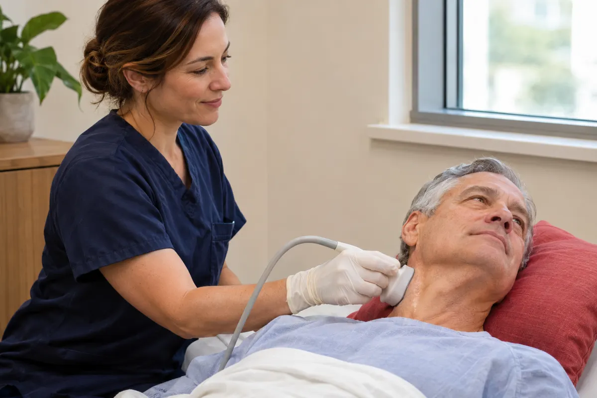
Place two fingers gently on the side of your neck, just below the angle of your jaw, and you can usually feel a strong, steady pulse. That pulse belongs to one of your carotid arteries, the two main vessels that carry blood from your heart up to your brain. Because they supply so much of the brain’s blood, the health of these arteries is closely tied to stroke risk.
A carotid ultrasound is a simple, non-invasive way to look at these arteries and the blood flowing through them. If your doctor has recommended one, or you are curious why it is part of some people’s heart and circulation check-ups, this guide explains what the test looks for, how it fits into stroke risk assessment and what the appointment is like at our clinic in Ajax.
Know the signs of stroke: act FAST
Before anything else, it is worth knowing how to recognize a stroke, because fast action matters. Heart & Stroke in Canada uses the word FAST to help people remember the most common signs:
- F – Face: Is it drooping?
- A – Arms: Can you raise both?
- S – Speech: Is it slurred or jumbled?
- T – Time: Call 911 right away.
If you or someone near you shows any of these signs, call 911 immediately, even if the symptoms seem to go away. Do not drive to the hospital yourself, and do not go to an imaging clinic. A carotid ultrasound is a planned, outpatient test. It is not a way to check for a stroke that is happening now, and diagnostic imaging clinics are not equipped to provide emergency care.
Symptoms that come and go, sometimes called a mini-stroke or transient ischemic attack (TIA), are also a warning sign and need emergency assessment. Call 911.
The carotid arteries in brief
You have two carotid arteries, one on each side of the neck. Each one rises from the chest and, partway up the neck, divides into two branches:
- the internal carotid artery, which carries blood into the skull to supply much of the brain
- the external carotid artery, which supplies the face, scalp and neck
The area where the artery divides, known as the bifurcation, is a spot where plaque tends to build up. It is one of the main areas examined during a carotid ultrasound.
How narrowing relates to stroke
Over time, fatty deposits, cholesterol, calcium and other material can collect inside artery walls. This buildup, called plaque, is part of a process known as atherosclerosis. It can happen in arteries throughout the body, including the carotids.
Plaque in a carotid artery can be linked to stroke in a couple of ways:
- Narrowing. As plaque grows, it can narrow the inside of the artery, a condition called carotid stenosis. Significant narrowing may reduce blood flow toward the brain.
- Fragments or clots. Small pieces of plaque, or a clot that forms on the surface of plaque, can break off and travel into smaller arteries in the brain, blocking blood flow there.
Not every stroke is caused by carotid artery disease, and many people with some plaque never have a stroke. But because carotid narrowing is one recognized contributor, imaging these arteries gives your physician valuable information when assessing stroke risk.
Why your doctor might order a carotid ultrasound
There are several reasons a health-care provider might request this test. Common ones include:
- Recent stroke-like symptoms, such as a TIA, once emergency assessment has taken place
- A bruit, which is a whooshing sound heard over the neck through a stethoscope, sometimes caused by turbulent blood flow
- Monitoring known narrowing that was found on a previous scan
- Follow-up after a procedure on the carotid arteries
- Risk factor assessment in some people with conditions that affect blood vessels
Risk factors for carotid artery disease overlap with those for heart disease and stroke in general. They include high blood pressure, high cholesterol, diabetes, smoking, older age, a family history of vascular disease, and existing heart or circulation problems. Your doctor decides whether a carotid ultrasound is useful in your situation. It is not a routine screening test for everyone.
What the scan actually measures
A carotid ultrasound combines two kinds of information.
Pictures of the artery wall
Greyscale ultrasound images show the walls of the carotid arteries and any plaque within them. The images can help describe where plaque is located and give a general sense of its extent.
Measurements of blood flow
Doppler ultrasound measures how fast blood is moving through the artery. This matters because blood speeds up as it passes through a narrowed section, much as water speeds up when you put your thumb over the end of a garden hose. By measuring blood flow speeds at different points, the exam helps estimate how much narrowing may be present.
Colour Doppler shows flow as colour on the screen, and spectral Doppler shows it as a waveform. You will likely hear the rhythmic whooshing sound of the Doppler signal during the scan, which is completely normal. For more on how the technology works, see our explainer on vascular ultrasound and blood flow imaging.
How it compares with other ways of imaging the neck arteries
Carotid ultrasound is often the first imaging test used to look at these arteries, but it is not the only one. Depending on the findings and the clinical question, your doctor or a specialist may sometimes recommend another type of imaging, usually arranged through a hospital.
- Ultrasound is non-invasive, uses no radiation and needs no injection. It gives real-time information about both the artery wall and blood flow. Its view is limited to the parts of the artery that can be reached from the surface of the neck.
- CT angiography uses X-rays and an injected contrast dye to create detailed cross-sectional pictures of the arteries, including portions higher up toward the skull.
- MR angiography uses magnetic fields and radio waves, sometimes with contrast, to produce detailed images of blood vessels without X-rays.
Each test has its own strengths. Ultrasound is well suited to an initial look and to monitoring over time, while the others may be used when more detail is needed or when planning a procedure. Which tests you need, and in what order, is a decision for your health-care team.
Why the result is only part of the picture
A carotid ultrasound tells your doctor about one set of blood vessels. Stroke risk, however, is shaped by many things at once, including blood pressure, heart rhythm, cholesterol, blood sugar, smoking history and age. Some strokes are related to heart rhythm problems or to small vessels deep in the brain rather than the carotid arteries. That is why your doctor considers your ultrasound results alongside everything else they know about your health, rather than treating the scan as a single answer.
What happens during the appointment
The exam is generally comfortable and requires little from you other than lying still.
- Clothing. Wear a top with an open or loose neckline. You will be asked to remove necklaces and earrings that might be in the way, so it can help to leave jewellery at home.
- Positioning. You lie on your back on an exam bed, often with your head tilted back slightly and turned away from the side being scanned.
- The scan. A clear gel is applied to your neck, and the technologist moves the transducer along the side of the neck from near the collarbone up toward the jaw. Both sides are usually examined.
- Your part. Stay relaxed and avoid talking or swallowing when asked, since movement can affect the images.
- Afterwards. The gel is wiped away, and you can return to your normal activities.
There are no needles and no radiation. The pressure of the transducer is light, though you may feel it more near the jaw. Let the technologist know if anything is uncomfortable.
Preparation
Preparation for vascular ultrasound varies by exam, and our team will confirm any instructions when your appointment is booked. Bring your health card and requisition, and allow a little extra time for parking and check-in.
Understanding results
After your exam, the findings are reported to the doctor who ordered the test. Your doctor will explain what the report means for you, which might include reassurance, changes to managing risk factors, follow-up imaging or a referral to a specialist. Decisions about treatment are made with your health-care provider, not at the imaging clinic.
It can help to bring a few questions to that follow-up conversation:
- Was any narrowing or plaque found, and on which side?
- Does this result change my overall stroke risk?
- Will I need another scan, and if so, when?
- Are there other tests, such as heart testing, that would be useful?
- What can I do day to day to look after my blood vessels?
Looking after your arteries
Whatever your results, many of the steps that support healthy arteries are the same ones recommended for heart health generally. Talk to your doctor about what applies to you, but common themes include knowing your blood pressure, cholesterol and blood sugar numbers, being physically active, not smoking, eating a balanced diet and taking medications as prescribed. If your doctor recommends heart testing, such as an echocardiogram or an electrocardiogram, these are offered through our cardiology team, Durham Cardiology, with a requisition.
Booking a carotid ultrasound in Ajax
Carotid ultrasound is part of our vascular ultrasound service, performed by certified technologists using high-precision equipment. Our clinic is at Unit 102, 300 Rossland Road East in Ajax, with free parking and ground-floor access, and we welcome patients from Ajax, Pickering, Whitby and across Durham Region.
If you have a requisition, call us at 905-426-3111 or request an appointment, and our team will confirm your booking and preparation. And please keep FAST in mind: for any sudden stroke signs, call 911 first.