
Achilles Tendon Injuries in Ajax: Ultrasound Basics
Learn how the Achilles tendon gets injured, which warning signs need the emergency department, and what Ajax patients can expect during an Achilles ultrasound.
· 8 min read
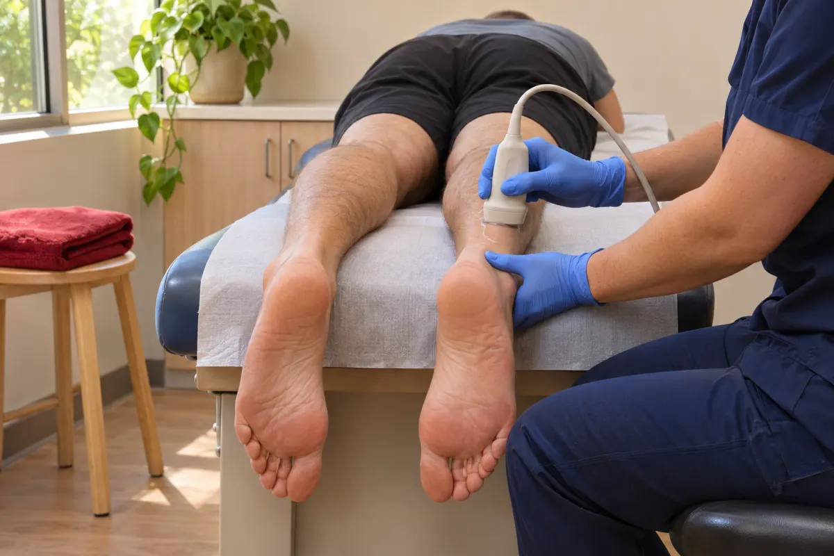
The Achilles tendon is the largest and strongest tendon in the body. It joins the calf muscles to the heel bone, and every time you walk, climb stairs, jump or rise onto your toes, it transmits the pull of those muscles to the foot. It handles that load thousands of times a day, mostly without complaint. When it does start to complain, though, it can bring an active routine to a halt.
This article covers the basics: how the tendon is built, the main types of Achilles problems, the signs that need emergency care rather than a booked test, and what an Achilles ultrasound involves. It is written for patients in Ajax and across Durham Region who have been referred for a scan, or who want to understand the test their provider has mentioned. It is not a substitute for an assessment by your own health-care provider.
The short version
- The Achilles tendon can be irritated gradually (tendinopathy), partly torn, or completely torn (rupture).
- A sudden pop at the back of the ankle, often with a feeling of being kicked, needs urgent assessment in an emergency department.
- Ultrasound is well suited to the Achilles because the tendon lies just under the skin and can be watched as the foot moves.
- Your provider interprets the scan alongside your symptoms and examination and decides on next steps with you.
How the tendon is built
The two main calf muscles, the gastrocnemius and the soleus, narrow into a single thick band that runs down the back of the lower leg and attaches to the back of the heel bone, the calcaneus. Around the tendon is a thin, slippery covering called the paratenon, which lets it glide. Near the attachment sit two small bursae, cushions that reduce friction between the tendon, the heel bone and the back of your shoe.
Providers often divide the tendon into regions when they talk about it:
- The muscle-tendon junction, higher up the calf, where muscle turns into tendon
- The mid-portion, a few centimetres above the heel, which is narrower and has a comparatively limited blood supply
- The insertion, where the tendon attaches to the heel bone
Different problems tend to favour different regions, which is one reason an ultrasound report usually describes exactly where along the tendon something was seen.
Types of Achilles problems
Mid-portion tendinopathy
This is a common pattern among runners, but it also affects people who are not especially active. The tendon becomes painful and often thickened a few centimetres above the heel. People may notice stiffness first thing in the morning or at the start of activity that eases as they warm up, then returns afterward. A tender, sometimes visibly swollen area along the tendon is typical.
Insertional tendinopathy
Here the pain is right at the back of the heel where the tendon meets bone. It can be aggravated by uphill walking, stairs and shoes with a firm heel counter that presses on the area. Bony changes or calcium deposits at the insertion are sometimes part of the picture, and these show on X-ray as well as ultrasound.
Paratenon irritation and bursitis
Sometimes the covering around the tendon or one of the nearby bursae becomes irritated, causing swelling and tenderness, with or without changes in the tendon itself.
Partial tears
A partial tear involves some of the tendon fibres but not the whole thickness. These may follow a sudden effort or develop in a tendon that has already been changing over time.
Complete rupture
A full tear of the tendon usually happens during a sudden push-off or landing, for example lunging for a ball, sprinting from a standstill or missing a step. It is often seen in people in mid-life who play sports intermittently, though it can happen at any age.
Warning signs that need emergency care
An imaging clinic is not the place to go first for a suspected rupture. Go to the nearest emergency department, or call 911 if you cannot get there safely, if you have:
- A sudden snap or pop at the back of the ankle or calf, often with the feeling that someone kicked you
- Sudden difficulty walking normally, pushing off with the foot, or rising onto your toes on that side
- A gap or dent you can feel in the tendon above the heel
- Calf pain with swelling, warmth and redness, particularly if you also have shortness of breath or chest pain
The last point is included because a painful, swollen calf can sometimes be caused by a blood clot rather than a tendon problem. Our article on blood clots and DVT ultrasound explains how vein scans are used. If you are unsure how urgent your symptoms are, Health811 provides free advice from a registered nurse in Ontario.
Factors linked to Achilles trouble
Providers consider several factors when they assess Achilles symptoms. None of them guarantees a problem, and many people with tendon pain have none of them.
- A recent jump in running distance, speed or hill work
- Returning to sport after a long break
- Tight calf muscles or changes in footwear
- Age, since tendons change gradually through adulthood
- Certain medical conditions and some medications, including a class of antibiotics called fluoroquinolones and corticosteroids, which have been associated with tendon problems
If you are concerned about a medication, do not stop it on your own. Talk to your doctor or pharmacist, who can weigh the benefits and risks for you.
Why ultrasound suits the Achilles
Ultrasound is one of the most commonly used tests for the Achilles tendon, and the reasons are practical.
- It is right under the skin. Sound waves can show the tendon in fine detail along its full length.
- It shows movement. The sonographer can watch the tendon as you gently point and flex your foot, which helps show how it glides and, where a tear is suspected, how the torn ends behave.
- It measures thickness. A tendon that is thicker than expected, or thicker than the other side, is a common finding in tendinopathy.
- It can show blood flow. A Doppler setting can show whether there is increased blood flow within the tendon, which is sometimes seen with tendinopathy.
- It allows side-to-side comparison, if that is part of the request.
- It does not use ionizing radiation.
An X-ray may be added if your provider wants to look at the heel bone, for example at bony changes near the insertion. If you want to understand how ultrasound looks at other tendons, ligaments and muscles, see our overview of musculoskeletal ultrasound for sports injuries. Achilles symptoms sometimes overlap with other heel problems, which our article on foot and heel pain imaging describes.
What happens during an Achilles ultrasound
The scan is straightforward and usually brief.
Positioning
You will lie face down on the exam table with your feet hanging just off the end. This lets the tendon relax and lets your foot move freely. If lying flat on your stomach is difficult, let the sonographer know, since there are other ways to position you.
Scanning
After applying gel, the sonographer moves a small transducer along the back of the lower leg, from the calf down to the heel, taking images both along the length of the tendon and across it. Expect gentle, steady pressure. If the tendon is very tender, mention it.
Movement
You may be asked to slowly point your toes and then pull them back toward your shin, or the sonographer may gently move your foot for you. Some sonographers also gently squeeze the calf during the scan. These movements are brief and help show how the tendon behaves in motion.
Clothing and what to bring
Wear shorts or loose trousers that roll up above the calf, and expect to remove shoes and socks. Bring your health card and requisition. No fasting or special preparation is usually needed, but follow any instructions given at booking. If you are wearing a walking boot or brace, the sonographer will guide you on whether and when to remove it.
After the scan
The report goes to the provider who ordered it. It may describe the tendon’s thickness, where along the tendon any changes were seen, whether a tear is partial or complete, and whether there is fluid or increased blood flow. Some of these words can sound alarming in isolation, so it is worth waiting to discuss them with your provider rather than searching each one online.
Decisions about rest, footwear, a structured exercise program, physiotherapy, a boot or referral to a specialist are made between you and your provider, based on the whole picture.
Booking an Achilles ultrasound
Rossland Diagnostic Clinic offers ultrasound by appointment at Unit 102, 300 Rossland Road East in Ajax, serving patients from Ajax, Pickering, Whitby and nearby. Parking is free, and the clinic has ground-floor access, so there are no stairs to manage on a sore leg.
When you have a requisition, call 905-426-3111 or request an appointment online, and our team will contact you to arrange a convenient time, including Saturday mornings.